인포그래픽 시각화 디자인
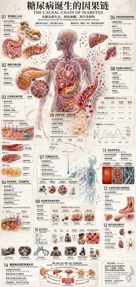
프롬프트
{
  "type": "medical infographic poster",
  "style": "highly detailed anatomical illustrations, clean structured layout, scientific diagrammatic style",
  "color_palette": "{argument name=\"color palette\" default=\"medical red, blue, beige, and anatomical flesh tones\"}",
  "language": "{argument name=\"language\" default=\"bilingual Chinese and English\"}",
  "header": {
    "main_title": "{argument name=\"main title\" default=\"糖尿病诞生的因果链\"}",
    "english_title": "{argument name=\"english title\" default=\"THE CAUSAL CHAIN OF DIABETES\"}",
    "subtitle": "从胰岛素失灵,到高血糖,到全身损伤"
  },
  "layout": {
    "centerpiece": "{argument name=\"central subject\" default=\"transparent human body showing circulatory system and internal organs\"}",
    "sections_count": 14,
    "sections": [
      { "id": "01", "title": "葡萄糖进入生命", "visuals": ["stomach and intestines"] },
      { "id": "02", "title": "胰腺与胰岛素", "visuals": ["pancreas", "beta cell"] },
      { "id": "03", "title": "正常胰岛素作用", "visuals": ["receptor signaling diagram", "muscle, liver, adipose icons"] },
      { "id": "04", "title": "胰岛素抵抗: 2型通路开始", "visuals": ["receptor blockage diagram", "7 lifestyle icons"] },
      { "id": "05", "title": "肝脏持续释放葡萄糖", "visuals": ["liver"] },
      { "id": "06", "title": "β细胞衰竭: 代偿到失败", "visuals": ["beta-cell decline line chart"] },
      { "id": "07", "title": "1型糖尿病分支", "visuals": ["autoimmune destruction diagram"] },
      { "id": "08", "title": "高血糖与血液化学", "visuals": ["blood vessel with glucose", "glucose indicators table", "glucose variability chart"] },
      { "id": "09", "title": "高血糖导致组织损伤", "visuals": ["4 pathways of damage diagrams"] },
      { "id": "10", "title": "急性代谢后果", "visuals": ["7 symptom icons"] },
      { "id": "11", "title": "微血管并发症", "visuals": ["eye", "kidney", "nerve cross-section"] },
      { "id": "12", "title": "大血管并发症与组织损伤", "visuals": ["heart", "brain", "diabetic foot"] },
      { "id": "13", "title": "器官系统长期代价", "visuals": ["text list"] },
      { "id": "14", "title": "糖尿病是调控系统失灵", "visuals": ["metabolic control flowchart"] }
    ],
    "footer": {
      "core_message": "核心信息 CORE MESSAGE"
    }
  }
}
한국어 번역
{
  "type": "의학 인포그래픽 포스터",
  "style": "매우 정교한 해부학 일러스트, 깔끔하고 구조적인 레이아웃, 과학 도해 스타일",
  "color_palette": "{argument name=\"color palette\" default=\"의학용 레드, 블루, 베이지, 해부학적 살색 톤\"}",
  "language": "{argument name=\"language\" default=\"중국어와 영어 이중 언어\"}",
  "header": {
    "main_title": "{argument name=\"main title\" default=\"당뇨병이 생기는 인과 사슬\"}",
    "english_title": "{argument name=\"english title\" default=\"THE CAUSAL CHAIN OF DIABETES\"}",
    "subtitle": "인슐린 기능 이상에서 고혈당으로, 그리고 전신 손상까지"
  },
  "layout": {
    "centerpiece": "{argument name=\"central subject\" default=\"순환계와 내부 장기가 드러난 투명 인체\"}",
    "sections_count": 14,
    "sections": [
      { "id": "01", "title": "포도당이 생명으로 들어오다", "visuals": ["위와 장"] },
      { "id": "02", "title": "췌장과 인슐린", "visuals": ["췌장", "베타세포"] },
      { "id": "03", "title": "정상 인슐린 작용", "visuals": ["수용체 신호전달 도해", "근육, 간, 지방 조직 아이콘"] },
      { "id": "04", "title": "인슐린 저항성: 2형 경로의 시작", "visuals": ["수용체 차단 도해", "7가지 생활습관 아이콘"] },
      { "id": "05", "title": "간이 포도당을 계속 방출하다", "visuals": ["간"] },
      { "id": "06", "title": "β세포 고갈: 보상에서 실패로", "visuals": ["베타세포 감소 꺾은선 차트"] },
      { "id": "07", "title": "1형 당뇨병 분기", "visuals": ["자가면역 파괴 도해"] },
      { "id": "08", "title": "고혈당과 혈액 화학", "visuals": ["포도당이 있는 혈관", "혈당 지표 표", "혈당 변동성 차트"] },
      { "id": "09", "title": "고혈당이 조직 손상을 일으키다", "visuals": ["4가지 손상 경로 도해"] },
      { "id": "10", "title": "급성 대사 결과", "visuals": ["7가지 증상 아이콘"] },
      { "id": "11", "title": "미세혈관 합병증", "visuals": ["눈", "신장", "신경 단면"] },
      { "id": "12", "title": "대혈관 합병증과 조직 손상", "visuals": ["심장", "뇌", "당뇨발"] },
      { "id": "13", "title": "장기 계통의 장기적 대가", "visuals": ["텍스트 목록"] },
      { "id": "14", "title": "당뇨병은 조절 시스템의 고장", "visuals": ["대사 조절 흐름도"] }
    ],
    "footer": {
      "core_message": "핵심 메시지 CORE MESSAGE"
    }
  }
}
템플릿 변수
치환 가능한 매개변수가 있으며, 대괄호 안이 기본값입니다
color palette = medical red, blue, beige, and anatomical flesh toneslanguage = bilingual Chinese and Englishmain title = 糖尿病诞生的因果链english title = THE CAUSAL CHAIN OF DIABETEScentral subject = transparent human body showing circulatory system and internal organs
출처
- 원작자
- hx831126
- 원문 출처
- 원문 보기 ↗
- 수록 출처
- freestylefly/awesome-gpt-image-2 · 67 · MIT